


Coksartrosis- This is a hip joint arthrosis.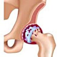 It grows gradually, for several years, exposed to development, can be one -sides and two sides.It is accompanied by pain and movement restrictions in the joints.In later stages, hip muscle atrophy and shortening of limbs are observed.Diagnosis is established based on clinical symptoms and radiographic results.In the early stages of coxarthrosis, conservative treatment.With joint destruction, especially in young and middle -aged patients, surgery (endoprosthetics) is indicated.
General information
Coksartrosis (osteoarthrosis or arthrosis of the hip joint deformation) is a degenerative-dystrophic disease.It usually grows at the age of 40.It may be due to various injuries and joint illnesses.Sometimes it happens for no apparent reason.Coksartrosis is characterized by progressive courses gradually.In the early stages, conservative treatment methods were used.In later stages, joint functions can only be restored.
In orthopedic and traumatology, coxarthrosis is one of the most common arthrosis.The high frequency of its development is due to significant burden on the hip joint and the prevalence of widespread congenital pathology - joint displacement.Women suffer from coksartrosis slightly more often than men.
The cause of coksartrosis
The main one (arises for unknown causes) and secondary (developed as a result of other diseases) the arthrosis of the hip joint is distinguished.
Secondary coksartrosis can be caused by the following disease:
- Displasia of the hip joint.
- Inblors of the weh.
- PERTES DISEASES.
- Aseptic necrosis of the thigh.
- Infectious lesions and inflammatory processes (for example, hip joint arthritis).
- Injuries (traumatic dislocation, hip fractures, pelvic fractures).
Coksartrosis can be either one -of -the -art or twice.With the main coxarthrosis, spinal cord injury (osteochondrosis) and knee joints (gonartrosis) are often observed.
Risk factors
Some of the factors that increase the likelihood of coxarthrosis development include:
- The burden of continuous improvement in the joints.It is often observed in athletes in people who are overweight.
- Blood circulation disorders, hormone changes, metabolic disorders.
- Spinal pathology (kyphosis, scoliosis) or stop (flat foot).
- Old and indecent age.
- An inactive lifestyle.
Coksartrosis itself is not inherited.However, certain features (metabolic disorders, features of framework and cartilage weakness) can be inherited by children from parents.Therefore, with the presence of blood clots suffering from coxarthrosis, the probability of the occurrence of the disease slightly increased.
Burdenomy
The hip joints are formed by two bones: ileum and femoral.The head of the thigh is articulated with the iliac bone acetabulum, forming a strange "hinge".During movement, acetabulum remains moving, and the femoral head moves in a variety of directions, ensuring flexibility, extension, abduction, carry and rotation hips.
During movement, the articular surface of the slide bone is unobtrusive compared to each other, thanks to the smooth, elastic and durable hyalin cartilage that covers the swivel cavity and thigh heads.In addition, Hyaline's cartilage performs a function that absorbs shock and is involved in the redistribution of loads during movement and walking.
In the joint cavity there is little fluid together, which plays the role of lubrication and provides hyaline cartilage.The joints are surrounded by strong and strong capsules.Above the capsule is a large femoral and gluteal muscle, which provides movement in the joints and, together with the Hyalin cartilage, as well as the shock absorber that protects the joints from injury with unsuccessful movements.
With coxarthrosis, the joint fluid becomes thicker and more viscous.The surface of the hyaline cartilage is dry, loss of smoothness, covered with cracks.Due to the roughness, the cartilage during movement is always injured in each other, causing their thinning and aggravating the pathological changes in the joints.When coxarthrosis lasts, the bones begin to change, "adjusting" to increased pressure.The metabolism in the joints is declining.In the final stages of coxarthrosis, severe atrophy of the muscles -The pain of the disease is observed.
Symptoms of coxarthrosis
The main symptoms of the disease include pain in the joint, inguinal, thighs and knee joints.Also, with chocolate, stiffness of movement and joint stiffness, gait disorders, deficiencies, hip muscle atrophy and shortening of the body on the side of the lesion.Characteristics of Coksartrosis are abduction restrictions (for example, patients are difficult to try on a chair).The presence of certain signs and their severity depends on the level of coxarthrosis.The first and most constant symptoms are pain.
OnCoksartrosis first stageThe patient complains of periodic pain, which occurs after physical activity (walking or walking).The pain is localized in the joints, less frequently in the thighs or knees.After rest, it usually disappears.Gait for coxarthrosis in the first stage is not broken, movement is fully preserved, no muscle atrophy.
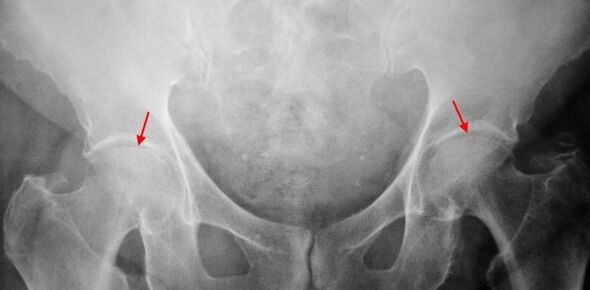
In the x -Ray patients suffering from coxarthrosis in the first stage, mild changes are determined: uneven narrowing of the joint gap, as well as bone growth around the external or internal edge of acetabulum if there is no change from the head and neck of the femur.
On2 -degree coksartrosisThe pain becomes more intense, often appears at rest, radiating to the thighs and thighs.After significant physical activity, patients with coksartrosis begin to drown.The amount of movement in the joints decreases: the abduction and rotation of the thighs are limited.
In image X -ray for the 2nd degree coxarthrosis, uneven narrowing of the joint gap (more than half of normal height) is determined.The femoral head is slightly turned upwards, defects and increases in size, and the contour becomes uneven.Bone growth with this level of coxarthrosis not only appears internally, but also on the outer edge of acetabulum and goes outside the cartilage.
OnCoksartrosis 3 degreesThe pain becomes continuous, worried about the patient not only during the day, but also at night.Walking is difficult, when moving, patients with coksartrosis are forced to use sticks.The amount of movement in the joints is limited, the muscles of the back, hips and lower legs are atrophied.Muscle weakness -The thighs that remove the thighs are the cause of the pelvic deviation in the front plane and shorten the limbs on the sore side.To balance the shortening, patients suffering from coksartrosis, while walking, tilt the body in a sore direction.As a result, the center of gravity shifts, the burden on the joints increases significantly.
On radiography for the 3rd degree coxarthrosis, sharp narrowing of the joint gap, significant thigh head development and double bone growth are detected.
Diagnostics
The diagnosis of coxarthrosis is based on clinical signs and additional study data, the main is radiography.In many cases, the X -Rays allows it to create not only the level of coxarthrosis, but also the cause of its occurrence.So, for example, an increase in the neck-dappyseal angle, scenes and leveling acetabulum indicate displacement, and changes in the form of femur proximal parts indicate that coksartrosis is a result of pertes or young epiphysiolysis.In radiography of patients with coxarthrosis, changes can also be detected indicate injury.
As other methods of diagnosis of instrumental coxarthrosis, CT and MRI can be used.Calculated tomography allows you to study pathological changes in detail by detailed bone structure, and magnetic resonance imaging provides an opportunity to evaluate disruption by soft tissue.
Differential diagnosis
First of all, coxarthrosis should be distinguished from gonarthrosis (osteoarthrosis of the knee joint) and spinal osteochondrosis.Muscle atrophy, which occurs at 2 and 3 stages of coxarthrosis, can cause pain in the knee joint, which is often expressed more brighter than pain in the damage area.Therefore, with patients' complaints about knee pain, clinical (examination, palpation, determination of movement amount) is a hip joint study, and if coxarthrosis is suspected, to direct the patient to radiography.
Pain for radicular syndrome (nerve compression) for osteochondrosis and some other spinal diseases can mimic pain with coxarthrosis.Unlike coxarthrosis, when rooting, pain occurs suddenly, after unsuccessful movements, sharp turns, weight lifting, etcPositive symptoms of tension are detected - severe pain when the patient tries to raise the member straight, lying on his back.At the same time, patients are free to take their feet to the side, while in patients with coksartrosis, abductions are limited.Keep in mind that osteochondrosis and coksartrosis can be seen at the same time, therefore, in all cases, a comprehensive examination of the patient is required.
In addition, chocolate is distinguished by trochanteritis (boot bursite) - aseptic inflammation in the gluteal muscle attachment area.Unlike coxarthrosis, the disease develops rapidly, within 1-2 weeks, usually after significant injury or physical activity.The intensity of the pain is higher than the coksartrosis.The limits of movement and shortening of members are not complied with.
In some cases, with unusual courses of disease or reactive arthritis, symptoms that resemble coxarthrosis can be observed.Unlike coxarthrosis, with this disease, the peak of pain falls at night.The pain syndrome is very intense, can decrease as it walks.Morning stiffness is a feature, which occurs shortly after waking up and gradually disappears within a few hours.
Coxarthrosis treatment
Pathological treatment is involved in the orthopedic traumatology.The choice of treatment method depends on the symptoms and levels of the disease.At 1 and 2 stages of coxarthrosis, conservative therapy is performed.During the coxarthrosis hunting, injection blocks, anti -non -—ssteroids (pyroxes, indomethacin, diclofenac, ibuprofen, and other) are used.Keep in mind that these groups are not recommended for a long time, as they can negatively affect the internal organizations and suppress Hyalin's cartilage ability to be restored.
To restore damaged cartilage for coksartrosis, funds from a group of chondroprotectors (chondroitin sulfate, cartilage extract, etc.) are used.To improve blood circulation and eliminate small vessels, vasodilating drugs (zinnarisine, nicotine acid, pentoxifillin, xanthinol nicotinate) are prescribed.According to the guidance, muscle relaxants are used (muscle relaxation drugs).
With stubborn pain syndrome, patients with coksartrosis may prescribe intra -articular injection using hormone drugs (hydrocortisone, triamcinolone, metrumor).Treatment with steroids must be carried out carefully.In addition, with coxarthrosis, local products are used - heating ointments that do not have a clear therapeutic effect, however, in some cases they relieve muscle cramps and reduce pain due to their "disruptive" actions.In addition, with coxarthrosis, prescribed physiotherapy procedures (glowing therapy, ultrasonic therapy, laser treatment, UHF, inductothermia, magnetotherapy), massage, manual therapy and therapeutic gymnastics.
Diet for coksartrosis has no free therapeutic effect and is only used as a way to lose weight.Losing weight allows you to reduce the burden on the hip joint and, as a result, facilitates the course of coksartrosis.To reduce the burden on the joints, doctors, depending on the level of coxarthrosis, may recommend walking with a stick or stick.
In later stages (with the 3rd degree coxarthrosis), the only effective treatment method is surgery -replacing joints that are destroyed with endoprosthesis.Depending on the nature of the lesion, either single -pocket (replacing only the head of the thighs) or two -pol (replacing both the thighs and the swivel cavity) can be used.
Endoprosthetics operation for coxarthrosis is performed in a planned manner, after complete examination, under general anesthesia.In the postoperative period, antibiotic therapy is performed.The stitches are released for 10-12 days, after which the patient is prescribed for the treatment of outpatients.After endoprosthetics, the recovery steps should be held.
In 95% of cases, surgical intervention to replace joints with coxarthrosis ensures complete recovery of limbs.Patients can work, actively move and play sports.The average service life of the prosthesis, subject to all suggestions, is 15-20 years.Subsequently, the second operation is required to replace the used endoprosthesis.