


Osteochondrosis is a disease of the spine, characterized by degenerative-dystrophic damage to the intervertebral discs, vertebral bodies and ligaments.
Osteochondrosis of the spine has a chronic progressive course.The disease is not felt for a long time, and symptoms appear only when complications arise.
According to statistics from the World Health Organization, 40-80% of the world's population suffers from osteochondrosis.
Among patients, people over 30 years old dominate.But, recently there is a trend towards the rejuvenation of osteochondrosis.Osteochondrosis ranks first among spine diseases in terms of disability among patients.
Brief anatomy of the spine
The spine performs the main functions - the spinal cord, support and movement, and also connects the head, shoulders and pelvic girdle.
The structural unit of the spine is the vertebra.
The 24 vertebrae are connected to each other by intervertebral discs, which are the body's shock absorbers.
The spine is divided into five parts: cervical, thoracic, lumbar, sacral and coccyx.
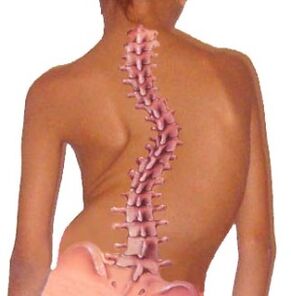
The normal shape of the spine is S-shaped.
The configuration of this organ makes it possible to distribute body weight and load evenly.
Structural and functional elements of the spinal column
A vertebra is a bony formation consisting of a body, arch and process.
The main load falls on the body of the vertebrae, so this is the largest part.
Important!Adjacent vertebral arches form the spinal canal - the container of the spinal cord, blood vessels, spinal nerve roots and adipose tissue.
LigamentsThe spine is represented by the posterior longitudinal ligament, which connects the vertebrae along the posterior surface, and the yellow ligament, the main purpose of which is to connect the vertebral arches.
Vertebral process.Vertebrae have 7 processes extending from the arches: spinous process, two transverse, two superior and two inferior articular processes.The ligaments and muscles of the spine are attached to the spinous process.Other processes form the intervertebral joints of the spine.
Intervertebral discsis a disc-shaped plate consisting of cartilaginous plate, annulus fibrosus and nucleus pulposus.Intervertebral discs connect adjacent vertebrae, providing mobility and stability to the spine.
Intervertebral jointsformed by the processes of two adjacent vertebrae.The main function of the intervertebral joints is to move the vertebrae relative to each other and provide flexibility to the spine.
Intervertebral foraminalocated on the lateral side of the spine and formed by the articular processes, bodies and pedicles of adjacent vertebrae.Spinal nerve roots exit through intervertebral foramina and blood vessels enter.
Spinal nerve- This is the part of the central nervous system that consists of nerve fibers.The spinal cord has three membranes - soft, arachnoid and hard.The dura spinal membrane consists of two sheets that connect and form a dural sac, filled with cerebrospinal fluid - cerebrospinal fluid.
Spinal nerve roots- This is the conductor of nerve impulses from the spinal cord to the internal organs and vice versa.Each spinal nerve root has autonomic, sensory and nerve fibers in its structure.
Paravertebral muscles- these are the muscles of the spine that support it and provide tilt and rotation of the body.
The functional unit of the spine isspinal motion segment, which consists of two adjacent vertebrae, intervertebral discs, ligaments and muscles.
Pathogenesis (mechanism of development) of spinal osteochondrosis
In the process of development, osteochondrosis passesfour levels:
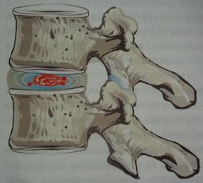
- First stage.Pathological changes do not go beyond the boundaries of the intervertebral disc.The nucleus pulposus dries up, which leads to a decrease in the height of the intervertebral disc.Fibrous rings cannot withstand the load - they crack and tear.
- Second stage.Due to the decrease in the height of the intervertebral disc, laxity of the ligaments and muscles of the spine occurs, which leads to instability of the spinal motion segment.The vertebrae can slip and move relative to each other.In this case, spondylolisthesis is formed.
- Third stage.The disease is growing.Intervertebral disc protrusion and intervertebral joint arthrosis, as well as uncovertebral joints, occur.
- Fourth stage.At this stage, the adaptive response is activated in the form of bone growth of the vertebral body (osteophytes).Therefore, the body tries to limit excessive vertebral mobility.Osteophytes with their sharp edges damage spinal nerve roots.Fibrous ankylosis of intervertebral discs and joints is formed, and the spine is immobile.The ankylosis stage is characterized by the loss of pain.
What leads to osteochondrosis?
Osteochondrosis of the backis a multifactorial disease where it is impossible to choose one specific cause.
The basis of osteochondrosis is a violation of microcirculation and metabolism in the spinal tissue, which can arise as a result of improper load distribution on the spine.
Factors that contribute to the development of osteochondrosis include the following:
- incorrect posture in childhood (scoliosis, kyphosis, kyphoscoliosis, stoop);
- back muscle weakness (inefficient spinal muscle corset);
- staying in one position for a long time (working on a computer, working in an office, doing handicrafts);
- improper weight lifting;
- physical inactivity and a sedentary lifestyle;
- metabolic pathology, especially lack of calcium, phosphorus, calcium, vitamins, magnesium, zinc;
- genetic predisposition to osteochondrosis;
- infectious diseases;
- frequent body hypothermia;
- chronic stress;
- hormonal imbalance;
- weight lifting;
- spinal cord injury;
- overweight and obesity.
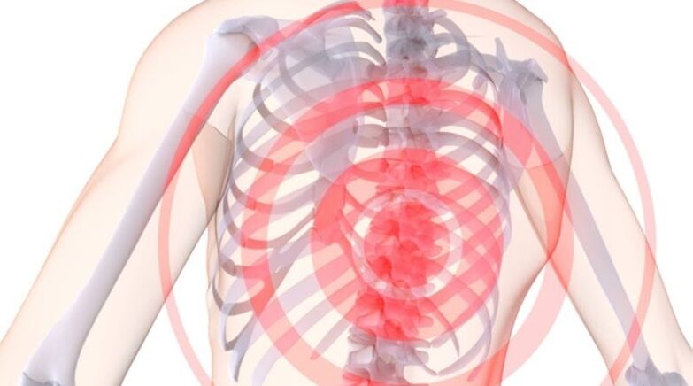
Symptoms of osteochondrosis
Chronic osteochondrosis can manifest itself with various symptoms.It all depends on the stage of the disease, the degree of damage to the spine and the presence of complications.
Clinically, the disease manifests itself when the degenerative-dystrophic process has reached the posterior part of the fibrous ring and the posterior longitudinal ligament, then the spinal nerve root is irritated, pinched and the flow of nerve impulses through it is disrupted.
At the same time, compression of the spinal cord and blood vessels occurs, which is indicated by reflex and compression syndrome.
Important!The pain syndrome in osteochondrosis occurs due to pinching of the spinal nerve roots in the intervertebral foramina by osteophytes, spasmodic muscles, and displaced vertebrae.
Osteochondrosis with its symptoms often mimics acute coronary syndrome, pleurisy, acute pancreatitis, hepatic and renal colic, acute appendicitis and adnexitis.
Therefore, it is important to carry out a comprehensive differential diagnosis of the disease to exclude life-threatening conditions.
Most commonsymptoms of osteochondrosis:
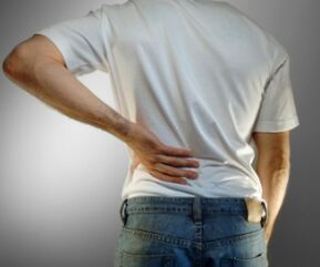
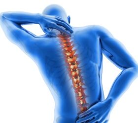
- pain in the neck, lower back, thoracic spine, which can be painful, throbbing or in the form of lumbago.The pain spreads to the head, upper and lower limbs, shoulder blades, heart, and stomach.The pain syndrome increases after physical activity, sneezing, laughing, coughing or staying in one position for a long time;
- sensory disturbancesdifferent parts of the body at the level of preservation of pinched nerves;
- crampsneck, back, upper and lower muscles;
- like a migraine headache;
- heartachein the joints of the limbs;
- increased fatiguefrom physical and mental labor;
- dizziness and loss of consciousnesswith a sharp turn of the head (vertebral artery syndrome);
- visual impairment(floating in front of the eyes or colored spots);
- decreased hearing acuity, tinnitus;
- pain in the heart;
- illalong the intercostal space;
- reduced blood supplythe upper and lower parts, indicated by the coolness of their skin;
- paresthesia– crawling, tingling and burning sensation in the spine;
- dry skin;
- sweating disorder;
- urinary disorders(dysuria, enuresis);
- decreased sexual desire, impotence.
Early diagnosis of osteochondrosis will greatly facilitate its treatment.
Methods for diagnosing osteochondrosis
A neuropathologist diagnoses osteochondrosis.If necessary, the patient can be referred to consult a cardiologist, gastroenterologist, orthopedic doctor, surgeon and others.
During the interview, it is necessary to determine exactly the nature of the complaint, when it arose, and what the patient associated with it.Be sure to check the medical history, profession of the patient, and whether a close relative has osteochondrosis.
Laboratory tests in this case are not informative.By conducting a biochemical blood test, you can pay attention to the level of calcium, phosphorus and other trace elements.
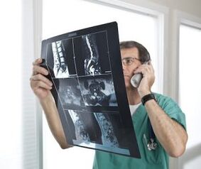
The main place in the diagnosis of osteochondrosis is occupied by instrumental methods, such as radiography of the spine, computed tomography and magnetic resonance imaging.
X-ray examination of the spine is the simplest, most accessible and very informative method for diagnosing osteochondrosis.
Radiography must be carried out in the direct and lateral projection of the desired part of the spine.Osteochondrosis is characterized by: a decrease in the height of the intervertebral disc, the presence of osteophytes, osteoporosis, and spinal deformity.
Myelography- This is an X-ray examination of the spine with the introduction of a contrast agent into the spinal canal.This method is dangerous because of the occurrence of an allergic reaction to the contrast.
Myelography allows us to study the internal structure of the spinal canal.This method is valuable for diagnosing Schmorl's hernia (intervertebral hernia).
Computed and nuclear magnetic tomography– this is a modern diagnostic method that visualizes the soft tissue and bone of the spine layer by layer.
This method is expensive, so it is used in severe cases, especially for the differential diagnosis of osteochondrosis and diseases with similar symptoms.
Since osteochondrosis is often disguised as a disease of the heart, lungs, pleura, stomach, intestines, kidneys, liver, there is a need for differential diagnosis.
For this purpose, the patient can be prescribed electrocardiogram, ultrasound examination of the heart and internal organs, blood test for troponin, ultrasound examination of blood vessels, chest radiography, electroencephalography and others.
Methods of treatment for osteochondrosis
Treatment of osteochondrosis canconservative and surgical.
Important!First of all, comprehensive conservative methods are used, and surgical treatment is used only in extreme cases.
Let's consider how osteochondrosis can be treated correctly.KconservativeTreatment methods for osteochondrosis can be listed:
- drug therapy;
- physical therapy;
- physiotherapeutic methods;
- manual therapy;
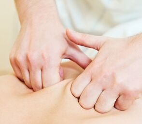
- massage;
- acupuncture.
Drug treatmentOsteochondrosis aims to relieve pain, relax muscles, relieve swelling of nerves and muscles, improve blood flow and conduction of nerve impulses.For this purpose, the following groups of drugs are used:
- nonsteroidal anti-inflammatory drugs;
- chondroprotectors, which includes a component of cartilage tissue.These drugs protect the vertebral cartilage and intervertebral disc from the negative effects of various factors;
- diureticwhich removes excess fluid from the body and relieves swelling of the spinal nerve roots and paravertebral muscles;
- muscle relaxantrelieves muscle spasms;
- drugs, improves metabolism and microcirculation in spinal tissue (vitamins B1, B6, B12, C, A and E);
- calcium supplements;
- hormonal drugs, which is prescribed when non-steroidal anti-inflammatory drugs are ineffective.
Therapeutic exercise– this is a dosed physical activity that can be done at home and at work for the treatment and prevention of osteochondrosis.
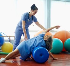
There are many sets of exercises for osteochondrosis.Prescribing exercise therapy and monitoring its implementation is carried out by a qualified specialist - physical therapist.
Thanks to properly selected exercise therapy, you can relieve pain, improve movement and blood supply to the spine, and stop the progression of the disease.
Physiotherapy treatmentosteochondrosis is carried out in special physiotherapy departments of hospitals, sanatoriums, and dispensaries by physiotherapists.
Physiotherapy methods include: electrophoresis, magnetic therapy, laser therapy, mud therapy, balneotherapy, ultraviolet exposure on the affected part of the spine, vibration treatment and others.
Manual therapy– this is a dosed manual effect on the spine to restore its mobility, eliminating displacement of vertebrae and intervertebral discs.
Manual therapy can only be performed by a certified chiropractor.
Massage and self-massagefor osteochondrosis, it is done to relieve muscle spasms, improve microcirculation in the paravertebral tissue and increase the mobility of the spine.
Acupunctureis a method of treating osteochondrosis in which thin needles are injected into active points.
Under the influence of needles in the body, endogenous opiates and cortisol levels increase, which have anti-inflammatory and analgesic effects.
Prevention of osteochondrosis
To maintain your health and your spine remains mobile until old age, follow some principles for the prevention of osteochondrosis:
- pay attention to your posture– always keep your back straight, don't slouch;
- selectcorrect postureto sleep;
- sit right at the table(shoulders relaxed, back straight, furniture should match your height);
- while staying for a long time in one position (working in the office, at the computer, sitting at handicrafts), try every 1-1.5 hoursdo some physical exercise, massage yourself back, or just get up and walk;
- distribute the load properlyon the spine when lifting and carrying various weights;
- wear orthopedic shoes;
- healthy sleepon a flat, hard to medium hard mattress.It is better to buy orthopedic mattresses and pillows.
Osteochondrosis of the spineis a chronic progressive disease that, unfortunately, cannot be cured.The effectiveness of treatment directly depends on its timeliness.
Do not self-medicate so as not to worsen your condition.At the first signs of osteochondrosis, contact a neurologist.